The Non-Invasive Cardiodiagnostics department at Phoenix Children’s, also known as the Echo Lab, is designed to provide testing for patients who have a diagnosis or a suspicion of having congenital heart disease or heart failure. Once a cardiologist makes a diagnosis of a congenital heart issue, heart disease, or wants to rule out any problems in the heart, our department performs the imaging tests that show the doctors the heart and how well it performs.
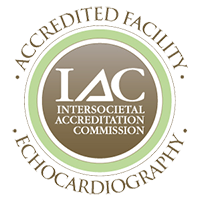
IAC Accreditation
Phoenix Children’s Echo Lab is proud to have earned the certification of Intersocietal Accreditation Commission for Pediatric and Adult Congenital Echocardiography, Fetal Echocardiography and Transesophageal Echocardiography.
Earning the IAC seal demonstrates that Phoenix Children’s has undergone the rigorous assessment by medical experts in pediatric and adult congenital echocardiography and meets the high standards set forth by the IAC to ensure quality patient care is being given.
The IAC accreditation is an ongoing review of our work for the technical quality of our images, the completeness of our studies, and the adherence to protocols and policies. Our studies are evaluated to ensure they meet the appropriateness criteria for ordering studies, that our results correlate with the results of other imaging studies and that our physician reports are regularly reviewed for completeness, accuracy and timeliness.


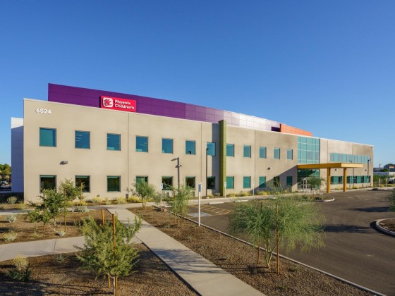
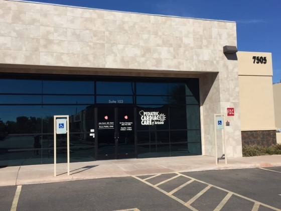
Phoenix Children's Specialty Care - Arrowhead Campus
6524 W. Sack Dr.
Glendale, AZ 85308
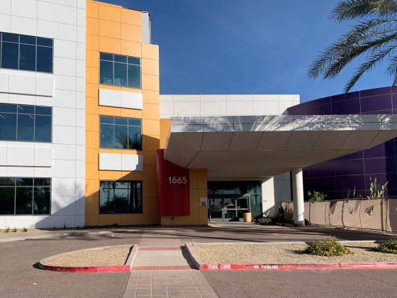
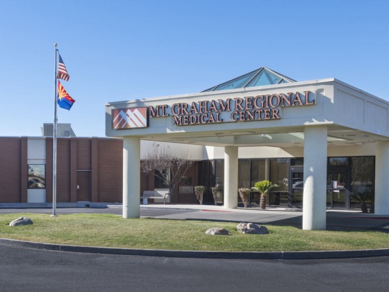
Phoenix Children's Specialty Care - Avondale Campus
1665 N. Avondale Blvd.
Avondale, AZ 85392