The Fetal Imaging Center at Phoenix Children’s uses state-of-the-art technologies for an accurate diagnosis and a personalized care plan. Advanced medical imaging can help your team of obstetric, maternal fetal medicine and pediatric specialists determine your baby’s condition. Your dedicated care coordinator will work with you to schedule any necessary imaging appointments at our main hospital, which features the latest in state-of-the-art imaging technology.

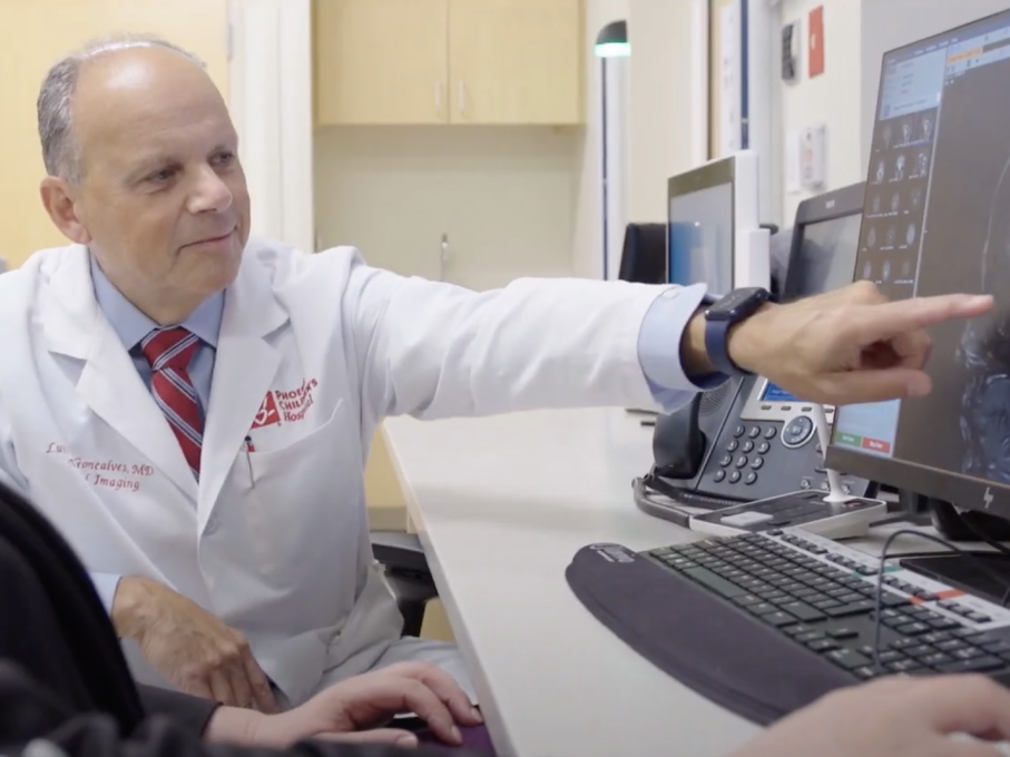
The caring and highly trained pediatric radiologists at Phoenix Children’s Fetal Imaging Center specialize in diagnosing conditions in unborn babies using advanced imaging technologies and techniques. By providing accurate diagnosis and personalized care planning, our goal is to better prepare you for what to expect during pregnancy, delivery and beyond.

Why Choose Us

Our team has performed thousands of imaging tests and brings decades of combined experience to every case. With radiologists who specialize in different areas of fetal imaging, we’re able to collaborate on complex cases to make the most accurate diagnosis possible.

Another advantage of choosing Phoenix Children’s is the seamless care your child receives before and after birth. We use prenatal images to guide delivery, plan treatment and support informed diagnoses after your baby is born. You’ll have us by your side from day one.

Advanced Technologies

The Fetal Imaging Center uses leading-edge technologies that produce the highest quality images of your unborn child, including:

  • MRI machines with the latest 3T and 1.5T magnets: Higher strength (3T) magnets help us examine certain problem areas in greater detail. 1.5T magnets produce better images of moving structures like the heart.
  • High-resolution 2D and 3D ultrasound equipment
  • Fetal echocardiograms 
  • 4D heart imaging technology
  • Dedicated 3D lab
  • Fast, low-dose computed tomography (CT) scanners

Our Imaging Tests

Fetal magnetic resonance imaging (MRI) is a safe, painless way to get clear pictures of your unborn baby. Unlike X-rays, MRIs don’t use radiation or pose risks to you or your child. Instead, they rely on magnets and radio waves to capture images. These scans can see through bones and fluids to create detailed 2D or 3D views.

Fetal MRIs happen after 20 weeks during your second or third trimester. We usually recommend one when an ultrasound raises questions that require further evaluation. They help us take a closer look at important areas – sometimes several at once – like the placenta, brain, heart and bones.

We use the information to understand your baby’s condition, plan for a safe delivery and decide on the best treatment.

Ultrasound uses reflected sound waves to produce an image of your unborn baby, the placenta and the surrounding amniotic fluid. It’s a safe way for imaging specialists to check on your baby’s development and look for any concerns.

At Phoenix Children’s, we use fetal ultrasounds in specific situations, often to complement MRI results or to evaluate certain high-risk conditions. These targeted scans help support the care you’re already receiving from your obstetrician or maternal-fetal medicine specialist.

Fetal computerized tomography (CT) scans use low-dose X-rays to create detailed, 3D images of your baby’s bones. We always use the lowest radiation necessary to minimize any risk.

Our radiologists only recommend CT scans in select cases, like for skeletal dysplasia or when other imaging doesn’t provide enough detail. They’ll carefully discuss this decision with you, your care team and referring provider before moving forward.