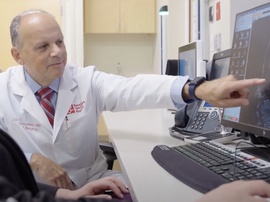
The Fetal Imaging Center at Phoenix Children’s uses state-of-the-art technologies for an accurate diagnosis and a personalized care plan. Advanced medical imaging can help your team of obstetric, maternal fetal medicine and pediatric specialists determine your baby’s condition. Your dedicated care coordinator will work with you to schedule any necessary imaging appointments at our main hospital, which features the latest in state-of-the-art imaging technology.
The caring and highly trained pediatric radiologists at Phoenix Children’s Fetal Imaging Center specialize in diagnosing conditions in unborn babies using advanced imaging technologies and techniques. By providing accurate diagnosis and personalized care planning, our goal is to better prepare you for what to expect during pregnancy, delivery and beyond.
What to Expect
When your physician sends an order to the Fetal Imaging Center at Phoenix Children’s, one of our registered nurse care coordinators will contact you to schedule your test. Your care coordinator will give you detailed instructions on what to expect on the day of your test.
It’s important to follow these instructions before your fetal imaging test:
- Do not wear jewelry or clothes with metal parts, such as snaps or zippers.
- Please arrive one hour before your test is scheduled, and bring a photo ID.
- Park in the Thomas garage, in front of the main hospital near the entrance.
Your physician may order one or more of the following tests:
If you will be having a fetal MRI, expect to be at the Fetal Imaging Center for three to four hours. These instructions will help you get started:
- Use the main entrance and sign in at the visitor’s desk. From there you will be directed to Radiology check-in.
- Take a seat in the waiting area, where a registration person will call you when they are ready.
- Tell the technician if you have any implanted medical devices, such as shunts, rods or nerve stimulators that are made of metal.
MRI scans usually take about one hour, but if your baby moves a lot, it could take longer or need to be repeated. The technologist will let you know if you need another test. Your MRI will proceed with these steps:
- You will lie on your back on the MRI table. If this is very uncomfortable, we can perform the MRI with you lying on your side.
- During the MRI, you will hear loud noises. We will give you earplugs to minimize the noise.
- You will have a button to push if you become very uncomfortable during the test.
- You may be asked to hold your breath at some point. This is because breathing movements can make the images blurry.
The goal is to get all the images the radiologist needs to make an accurate diagnosis. In some cases, an ultrasound will be done after the MRI to make sure there is enough information.
The radiologist will examine the images as they are taken, and then meet with you after the test is completed to review the preliminary images. After meeting with you, your radiologist will examine the images in more detail and take measurements.
The radiologist will then call your referring physician with the results, and also send a report. You can call your physician in a couple of days to ask about the best way to get the final results.
Fetal ultrasound is done over the abdomen (transabdominal ultrasound) or can be done through the vagina (transvaginal ultrasound) in special cases.
For a transabdominal ultrasound, wear loose-fitting clothing so you can lift it to expose your abdomen. Your test will proceed with these steps:
- You’ll lie on an exam table with your abdomen exposed.
- A technician will apply a warm gel to your abdomen. This helps transmit the sound waves to your uterus.
- The technician may ask you to move into several positions to get a better view of the fetus.
- After the procedure, you can wipe off any remaining gel.
For a transvaginal ultrasound, you’ll be asked to undress from the waist down or put on a hospital gown. You’ll also empty your bladder before the test. Transvaginal ultrasounds proceed with these steps:
- You’ll like on an exam table and put your feet in stirrups, similar to a gynecological exam.
- A transducer, which produces sound waves that create the images, is inserted into your vagina. It is covered with a plastic sheath and lubricated with warm gel.
The radiologist will analyze the images and send a report on your test to your physician, who will share it with you.
This test usually takes about 30 minutes overall, with an actual scanning time of just a few seconds. It proceeds with these steps:
- The radiologist will begin by doing a brief ultrasound to find the top and bottom of your uterus.
- With these borders marked, you’ll lie on a table that moves into a large, doughnut-shaped device.
- The CT scan captures the images of the fetus in seconds.
After your CT scan, the radiologist will examine the images of your baby’s bones and send a report to your physician, who will let you know the findings and diagnosis.
Advanced Technologies
The Fetal Imaging Center uses state-of-the-art technologies that produce the highest-quality images of your unborn child, including:
- MRI machines with the latest 1.5T and 3T magnets – the higher strength (3T) magnets allow us to examine certain problem areas in greater detail, while the 1.5 magnets produce better images of moving structures such as the heart
- High-resolution 2D and 3D ultrasound equipment
- Fast, low-dose computed tomography (CT) scanners
- Fetal echocardiography
Our 3D images are processed in our dedicated 3D lab.
Our Imaging Tests
Fetal magnetic resonance imaging, or fetal MRI, uses magnets, radio waves and a computer to make 2D or 3D pictures of your unborn baby. MRIs do not use X-rays and have no known side effects for your or your baby. These painless tests penetrate past bones and fluids in the body to produce clear, detailed images.
Fetal MRIs are usually done in the second or third trimester, after 20 weeks of pregnancy. They can reveal conditions of the placenta, brain, nervous system, heart, bones and other areas.
MRI provides information to help your doctor diagnose your unborn child’s condition, predict how your delivery will proceed and determine therapies for your baby.
Ultrasound uses reflected sound waves to produce an image of your unborn baby, the placenta and the amniotic fluid around the baby. Fetal ultrasound is a safe way for imaging specialists to view your baby and check for problems. The Phoenix Children’s radiology team may use this technique along with fetal MRI, since these tests can provide additional information when used together.
Fetal computerized tomography (CT) scans use low-dose X-rays to produce extremely accurate, 3D images of the unborn baby’s skeleton. Radiologists at Phoenix Children’s use this technology to examine the baby’s bones in special circumstances when an ultrasound or MRI test would not provide enough information for a definitive diagnosis.
Accurate Diagnosis for a Better Plan
With an accurate diagnosis, you and your family can see one or more of Phoenix Children’s nationally renowned pediatric specialists — at a location close to your home.
We collaborate closely with maternal-fetal medicine specialists, obstetricians and families to offer the highest level of care for you and your child.
Using clear, high-resolution images, our expert fetal and pediatric radiology specialists are committed to providing your baby’s care team with imaging results and expert insights needed to drive accurate diagnoses, treatment planning, parental counseling and decision making — without high radiation doses.
And, our imaging services are specifically designed with your comfort and your baby’s safety in mind.
Related Services
Locations
Phoenix Children's Specialty Care - Arrowhead Campus
6524 W. Sack Dr.
Glendale, AZ 85308